Last week we enjoyed a favourite blog post from the past on Creating Figures for Your Manuscript. It seems appropriate to review the corresponding post on Figure Legends this week. I hope you’ll find this information helpful.
According to my Twitter feed, a number of researchers are unsure of how to approach figure legends. I actually find figure legends fairly formulaic and easy to write.
As a first step, I recommend browsing through several recent issues of the journal where you’ll be submitting your paper. Look at the figures and how the captions are written. The structure should be fairly consistent across all papers, regardless of the topic, methodology, or type of image.
A quick disclaimer here: Studies published in some of the very high impact journals, such as Nature and Proceedings of the National Academy of Sciences, tend to have complex, multi-part figures which therefore require more complex figure captions. We will not be reviewing those here, but my tip from above applies: look at lots of sample articles. Pick papers that used techniques similar to those in your study, and model your figure legends accordingly.
Figures should be able to stand independently from the rest of the paper, so it’s important the legend includes all of the essential information. Nevertheless, the legends should be concise.
The bulk of scientific and medical journals follow a rather standard formula for figure legends.
Single Image Figure:
- Identify the type of image. Examples: radiograph (anteroposterior or lateral view), bar graph, flow chart, survivorship curve, electron micrograph.
- Describe the sample, patient, or cohort(s). For a sample, describe the cell type, protein or molecule, the line name, and any other identifiers or qualifiers. For a patient, include age, sex, left or right side (if an image of a bilateral appendage), the condition shown in the image, and any relevant comorbidities. For a cohort, define the type of patient included, the condition or procedure they have in common, and the sample size (n = ?).
- Define the point in time that the image was taken. Examples: preoperative, intraoperative, 6 months postoperative, at mean 18.5 ± 2.8 months follow-up.
- Define the procedure that was performed, highlighting only the most essential data. Examples: radioimmunoassay, antibodies and markers used, type of surgery, relevant hardware details.
- Point out any particular items of interest in the figure.
- Define any arrows, asterisks, colour coding, magnification, and measurement bar size.
- Define any acronyms used in a graph.
For multiple image figures, follow the same steps as above. First, describe all of the components from steps 1 through 4 that are common for all of the images within the figure plate, such as the type of image, point in time, patient or sample. Then list the individual images [ (a) … (b) … ] and describe what differentiates each of the images from the others.
Here are example figure legends for the 2 figures shown:
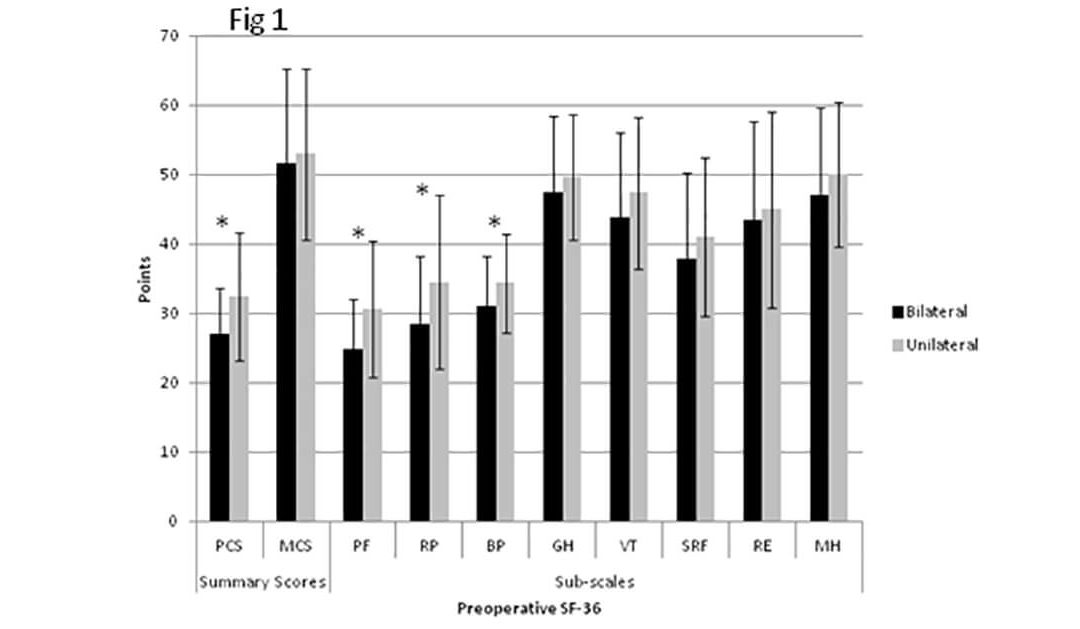
Figure 1: Bar graph showing mean preoperative SF-36 scores for patients with unilateral ankle arthritis (n=106) and bilateral ankle arthritis (n=52). Error bars indicate standard deviation. PCS = Physical component summary; MCS = Mental component summary; PF = Physical functioning; RP = Role-physical; BP = Bodily pain; GH = General health perceptions; VT = Vitality; SRF = Social role functioning; RE = Role-emotional; MH = Mental health. *Significant difference between unilateral and bilateral cohorts (p<0.05).
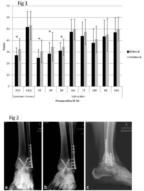
Figure 2: Radiographs of left ankle of 71-year-old male with isolated end-stage post-traumatic ankle arthritis, 52 weeks following ankle fusion, with a nonunion of the fusion site, as depicted by persistence of the joint space: (a) anteroposterior, (b) oblique, and (c) lateral views.
When you have your “final” figure and legend, ask another investigator, postdoc, or grad student who is not familiar with the details of your research to review it. If they don’t understand something, you can add the necessary details.
Was this tip helpful?
I welcome comments, as well as suggestions for future weekly writing tips.
I appreciate you taking the time to create this post. It has long been extremely helpful to me in fact. Appreciate it.
You can follow me on twitter @Dagmar__Gross (that’s with 2 underscores)